

Routine ultrasound
The results of ultrasound testing provide you and your health care provider with critical information about you and your baby. The most accurate way to “date” your pregnancy is with an early ultrasound. The Society of Obstetricians and Gynaecologists of Canada recommends that all women have two ultrasounds: one “dating” ultrasound at 11-14 weeks and one “anatomic” ultrasound between 18-20 weeks. See below for descriptions of the information gathered from these two types of ultrasounds.
What is an ultrasound?
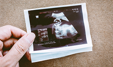 Ultrasound uses high-frequency sound waves to create images of the inside of the body. The technique does not use any radiation. It is safe, painless and relatively quick test that usually takes around 30 minutes. The technician will put a warm gel on your abdomen and use a scanning device to get the ultrasound images. Sometimes the ultrasound must be done through the vagina; this procedure may be uncomfortable, but is not considered painful. Ultrasound is not used to diagnose pregnancy, but is used to date a pregnancy and assess numerous health aspects of the fetus and mother.
What is my health care provider looking for on the ultrasound pictures?
Your ultrasound(s) gives important information on many aspects of your pregnancy. These include:
- The number of babies
- The gestational age of the baby
- The growth and development of the baby
- Whether the baby is the right size for his/her age
- How the baby’s internal organs are developing
- The location of the placenta
- The possibility of a genetic or other abnormality
- The possibility of an ectopic or molar pregnancy
- What position the baby is in
- How much fluid is around the baby
- How active the baby is
What is a “dating” ultrasound?
A dating ultrasound gives an accurate estimate of how far along you are in your pregnancy. Many women are uncertain of exactly when conception happened. Ultrasound can tell you how many weeks pregnant you are, based on the size of your fetus. Dating ultrasounds are most accurate when they happen between 7-12 weeks, and are calculated by measuring the fetus’ crown-rump length. This generally predicts the expected date of birth within 5 days.
Knowing where you are in your pregnancy is important for your prenatal care, all the way from your first trimester, through to labour.
What is an “anatomic” ultrasound?
This ultrasound is usually offered in the second trimester, between 18 and 22 weeks of gestation. As the name suggests, this type of ultrasound looks at your baby’s anatomy. The number of fetuses, gestational age, and location of the placenta will be assessed. The ultrasonographer will take many measurements of your baby to screen for any abnormalities. Images will be captured to view the development of your baby’s brain, face, heart, spine, chest, major organs, feet, and hands. The position of your placenta and the vessels in the umbilical cord will be examined. The amount of fluid around the baby will be measured, and your cervix, uterus, ovaries, and bladder will be assessed for any abnormalities. If you are at high risk for fetal abnormalities, anatomic ultrasound can be performed during early pregnancy (11-16 weeks). If the baby’s sex is determined during the ultrasound, you have the right to request this information.